Annotation
- Introduction
- The Evolution of AI in Medical Imaging
- Building the Diagnostic Neural Network
- AI as Collaborative Medical Support
- Practical Implementation Workflow
- Clinical Advantages and Operational Benefits
- Technical Architecture and Deployment
- Future Directions and Ethical Considerations
- Pros and Cons
- Conclusion
- Frequently Asked Questions
AI X-Ray Analysis: How Neural Networks Revolutionize Medical Imaging Diagnosis
AI-powered X-ray analysis uses neural networks to revolutionize medical imaging, enhancing diagnostic accuracy, efficiency, and patient care through
Introduction
Artificial intelligence is fundamentally reshaping healthcare delivery, with medical imaging standing at the forefront of this technological revolution. AI-powered X-ray analysis represents one of the most promising applications, combining sophisticated neural networks with traditional radiology to enhance diagnostic accuracy and efficiency. This comprehensive guide explores how these systems work, their practical implementation, and the transformative impact they're having on medical professionals and patient care worldwide.
The Evolution of AI in Medical Imaging
The integration of artificial intelligence into medical imaging marks a significant advancement in diagnostic technology. Unlike traditional methods that rely solely on human interpretation, AI systems leverage deep learning algorithms to analyze X-ray images with remarkable precision. These neural networks are trained on vast datasets containing thousands of annotated medical images, enabling them to recognize patterns and anomalies that might escape even experienced radiologists. The technology has evolved from simple pattern recognition to complex diagnostic assistance tools that can process images in seconds rather than minutes.
Modern medical image analysis systems utilize convolutional neural networks (CNNs) specifically designed for visual data processing. These networks can detect subtle variations in tissue density, identify foreign objects, and flag potential abnormalities with consistent accuracy. The training process involves feeding the system labeled examples of both normal and pathological findings, allowing the AI to learn the distinguishing features of various conditions.
Building the Diagnostic Neural Network
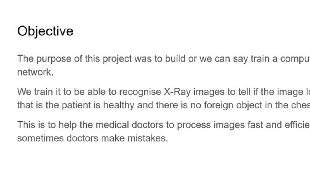
The core objective in developing AI for X-ray analysis involves creating a sophisticated neural network capable of distinguishing between healthy patients and those requiring medical attention. This system specifically targets chest X-rays, training to identify foreign objects and other abnormalities that might indicate underlying health issues. The development process begins with data acquisition from reputable sources like Kaggle, where thousands of anonymized medical images provide the foundation for machine learning.
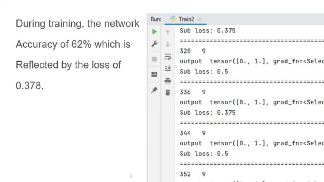
Using the PyTorch framework, developers construct neural networks with multiple layers that progressively extract and analyze features from input images. Each layer specializes in recognizing different aspects of the X-ray, from basic edges and shapes to complex anatomical structures. The training phase involves adjusting millions of parameters until the network achieves optimal performance in classification tasks.
AI as Collaborative Medical Support
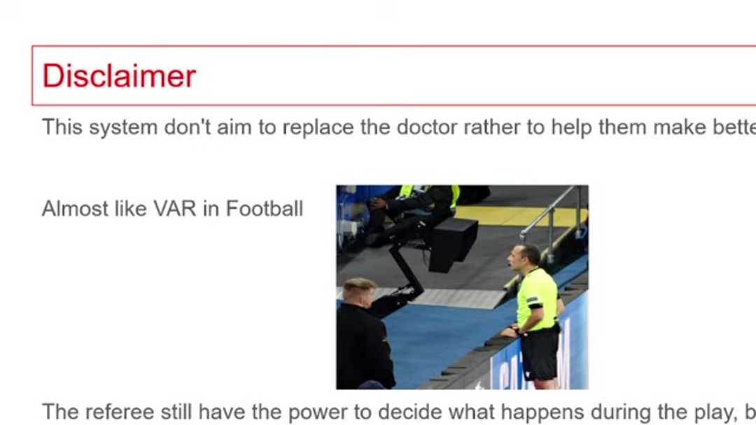
It's crucial to understand that AI in medical imaging serves as a collaborative tool rather than a replacement for human expertise. Think of these systems as digital assistants that provide second opinions, similar to video assistant referees in sports. They offer additional perspectives and flag potential concerns, but the final diagnostic decision remains with qualified medical professionals. This collaborative approach leverages the strengths of both human intuition and machine precision.
The system operates as a safety net, helping radiologists avoid oversight errors that can occur during high-volume work periods or when dealing with subtle findings. By providing consistent, objective analysis, AI assistants enhance rather than replace clinical judgment. Medical professionals maintain ultimate authority while benefiting from the AI's rapid processing capabilities and pattern recognition skills that complement their own expertise.
Practical Implementation Workflow
Implementing AI X-ray analysis follows a structured workflow designed for clinical efficiency. Users begin by accessing the web platform at x-ray-processing.web.app, where they can register and log into the system. The interface guides medical professionals through a straightforward process of uploading compatible X-ray images, typically in 1024x1024 pixel grayscale format that optimizes processing accuracy.
Once uploaded, the system processes the image through its trained neural network, analyzing pixel patterns and comparing them against learned representations of normal and abnormal findings. The results display as probability percentages indicating the likelihood of "Finding" or "No Finding," providing quantitative data to support diagnostic decisions. This streamlined approach significantly reduces waiting times for preliminary assessments.
Clinical Advantages and Operational Benefits
The implementation of AI in radiology departments brings multiple advantages that extend beyond simple automation. These systems can process images continuously without fatigue, maintaining consistent performance standards regardless of workload volume. The technology also demonstrates particular strength in identifying early-stage conditions that might present with subtle radiographic signs, potentially enabling earlier interventions and improved patient outcomes.
From an operational perspective, healthcare technology incorporating AI helps optimize resource allocation by prioritizing cases based on algorithmic risk assessment. This allows radiologists to focus their expertise on complex cases while routine screenings receive automated preliminary analysis. The systems also provide valuable training tools for medical students and junior radiologists, offering immediate feedback and comparison against expert-level interpretations.
Technical Architecture and Deployment
The underlying technology stack for AI X-ray analysis combines several robust frameworks to ensure reliability and scalability. The backend typically utilizes Django, a Python-based web framework known for its security features and rapid development capabilities. This interfaces with frontend applications built using Angular, providing responsive user experiences across different devices and platforms.
Deployment involves cloud hosting solutions like Digital Ocean, which offer the computational resources necessary for image processing while maintaining data security standards. MySQL databases manage patient information and analysis results, ensuring organized storage and retrieval of historical data for comparison and tracking purposes. This architecture supports the heavy computational demands of neural network inference while maintaining clinical workflow efficiency.
Future Directions and Ethical Considerations
As AI technology continues advancing, we can expect even more sophisticated applications in medical imaging. Future developments may include multi-modal analysis combining X-rays with other imaging techniques, predictive analytics for disease progression, and personalized treatment recommendations based on algorithmic findings. However, these advancements must be balanced with careful consideration of ethical implications, including data privacy, algorithmic transparency, and appropriate implementation protocols.
The medical community continues developing standards and guidelines for AI integration, ensuring these powerful tools enhance rather than complicate patient care. Ongoing research focuses on improving model interpretability, reducing biases in training data, and establishing clear protocols for human oversight in AI-assisted diagnostics.
Pros and Cons
Advantages
- Significantly accelerates diagnostic turnaround times
- Reduces human error through consistent analysis
- Operates continuously without fatigue or breaks
- Helps identify subtle patterns human eyes might miss
- Provides valuable second opinions for complex cases
- Optimizes radiologist workflow and case prioritization
- Supports medical education and training programs
Disadvantages
- Requires substantial initial investment in infrastructure
- Dependent on large, diverse datasets for training
- Potential algorithmic bias based on training data
- Limited explainability of AI decision processes
- Raises data privacy and security concerns
Conclusion
AI-powered X-ray analysis represents a significant step forward in medical technology, offering enhanced diagnostic capabilities while supporting healthcare professionals in their critical work. These systems combine the pattern recognition power of neural networks with human clinical expertise, creating collaborative diagnostic environments that benefit both medical providers and patients. As the technology matures and addresses current limitations around explainability and data requirements, we can expect even broader adoption across healthcare systems worldwide. The future of medical imaging lies in this harmonious integration of artificial and human intelligence, working together to improve diagnostic accuracy, efficiency, and ultimately patient outcomes.
Frequently Asked Questions
How accurate is AI in analyzing X-ray images compared to human radiologists?
AI algorithms achieve accuracy levels comparable to experienced radiologists for specific tasks, particularly in detecting obvious abnormalities and foreign objects. However, human experts still outperform AI in complex cases requiring contextual understanding and clinical correlation.
What training data sources do medical AI systems use?
Medical AI systems primarily use anonymized datasets from platforms like Kaggle, research institutions, and hospital partnerships. These datasets contain thousands of labeled medical images with verified diagnoses for supervised learning.
Can AI completely replace radiologists in X-ray interpretation?
No, AI serves as a support tool rather than replacement. It assists with initial screening and flagging potential issues, but final diagnosis and treatment decisions require human clinical judgment, experience, and patient context.
What technical frameworks are used for medical AI development?
PyTorch and TensorFlow are popular frameworks for building neural networks. Deployment typically involves Django backends, Angular frontends, and cloud hosting platforms like Digital Ocean for scalable medical applications.
What infrastructure is needed for AI X-ray analysis systems?
AI X-ray analysis requires robust computational infrastructure, including cloud hosting for processing, databases for storage, and secure networks to handle medical data, often built with frameworks like Django and Angular.
Relevant AI & Tech Trends articles
Stay up-to-date with the latest insights, tools, and innovations shaping the future of AI and technology.
Grok AI: Free Unlimited Video Generation from Text & Images | 2024 Guide
Grok AI offers free unlimited video generation from text and images, making professional video creation accessible to everyone without editing skills.
Top 3 Free AI Coding Extensions for VS Code 2025 - Boost Productivity
Discover the best free AI coding agent extensions for Visual Studio Code in 2025, including Gemini Code Assist, Tabnine, and Cline, to enhance your
Grok 4 Fast Janitor AI Setup: Complete Unfiltered Roleplay Guide
Step-by-step guide to configuring Grok 4 Fast on Janitor AI for unrestricted roleplay, including API setup, privacy settings, and optimization tips